1000+ types of tissue microarray | Biocoreusa corp
  888-899-6266       info@BioCoreUSA.com   
0 item(s) $0.00 
 
 
Promotions: 

Buy one get one free trial slide. 
Code: FREETRIAL 


Selected Tissue MicroArrays with IHC marker results and photos are now available. For detailed information, please clicking HERE

We offer H&E and IHC high-resolution scanning images that allows customers to see a 100x-400x microscope image, please contact us for require

 
Bestellers
 
$255.00
Prostate disease spectrum tissue array,...
$395.00
Pancreas cancer tissue array with normal...
$255.00
PD-L1 tested non-small cell lung cancer...

Tissue Array

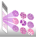
BioCoreUSA provides normal, malignant or metastatic tissue array or tissue microarray (TMA) which can be utilized to validate clinical relevance of potential biological targets in the development of therapeutics, diagnostics and study new protein markers and genes. TMA contains multiple tissue samples with DNA,RNA and protein. The advantage of TMA lies in the small size, and the tissue morphology and genetic characteristics are similar in the serial TMA tissue sections. Therefore, it provides a high-throughput methods for the fast analysis of molecular markers associated with disease diagnosis, prognosis and therapeutics in patients. BioCoreUSA also provides custom tissue array as well as the immunohistochemistry (IHC) staining service.

Tissue Bank/Tissue Sections

Tissue Bank provides whole tissue sections of paraffin embedded and formalin fixed tissue which are great for localizing DNA, RNA and protein markers. The tissue sections were fixed by formalin less than 24 hours, processed and sectioned with 4-5 μm thickness, and mounted on the positive charged SuperForst glass.

Services

Tissue Array Creation
Custom tissue microarray (TMA) construction with customer's tissue blocks or tissues
The Immunohistochemistry (IHC)
Services are provided to customers with specific needs for the tissue array experiments by IHC. 

DST244C CK18 FMG08013e_Ki-67 KIN481a_CK5_6 XHN483_P40
Histology Services
Paraffin tissue block sectioning, H&E staining. 
Image Scanning
Tissue section or tissue array core image digital capture using Imaging Scanning System from leading Microscopic Scanner company. 
Pathology Report
Professional pathology report including, pathology review, quantification of positive staining of the IHC as well as the scoring and location of positive staining manually by experienced pathologists in-house


BioCoreUSA Corporation
6024 Ridge Ave, Suite 116-236
Philadelphia PA 19128
United States
 
CONTACT US
Email:
Customerservices@biocoreusa.com
Info@biocoreusa.com
TEL: 888-899 6266
SUPPORT
Delivery
Privacy Policy
Terms & Conditions
FAQs